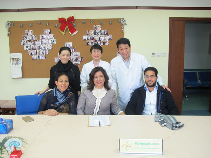
Luis Cedeno-Retinitis pigmentosa-(Dominican)-Posted on Mar.2nd, 2015
 Name: Luis Cedeno
Sex: Male
Country: Dominican Republic
Age: 28 years
Diagnosis: Retinitis pigmentosa
Date: Feb. 5th, 2015
Days Admitted to Hospital: 15 days
Before treatment:
At the age of 10, the patient got diminution of vision with no reason. At first, it did not affect the quality of his daily life activities. His illness became worse day by day, especially his left eye. At the age of 15, he was diagnosed with “Retinitis pigmentosa” after examination. But he didn’t receive any treatment. 5 years ago, his vision turned more serious too. 4 years ago, he had right eye surgery to improve his condition, but the surgery didn’t offer any good effect. He could only see object’s movement and have light sensitivity. His has tubular visual field of his left eye. His correct vision was 0.5 in the distance of 3 meters. He almost lost his vision of right eye. He could see fingers shaking in the distance of 10cm, but he couldn’t distinguish them. He wants to have a better treatment, so he came to our hospital and he was diagnosed with retinitis pigmentosa.
He was in good spirit. His sleep, eating, urination and excrement were good.
Admission PE:
Bp: 114/68mmHg; Hr: 70/min, Br: 18/min. He’s growth and nutrition was normal. His skin mucosa was without hemorrhaging spots or yellow stains. His thorax was symmetrical. The respiration of both lungs was clear, without moist rales. The heart sound was strong, and the cardiac rhythm was regular. There was no obvious murmur in the valves. The abdomen was flat and soft, with no masses. The liver and spleen were normal.
Nervous System Examination:
He was alert and his speech was clear. His memory, orientation and calculation ability were normal. The diameter of both pupils was 3.0mms and both pupils were sensitive to direct and indirect light reflex. His left eye was tubular visual field. 3 meters standard visual acuity chart: left eye corrected visual acuity 40/100; visible to the naked eye from 1 meter distance was 8/100. Color sense of right eye partly disappeared. His left eye had light sensitivity. He could see finger wave in front of 10cm, but he couldn’t see the objects’ shape. He lost his color sensitivity. Through use of an ophthalmoscope: right eye fundus was light pink. The color of retina in right eye was light yellow with a white tint, there were a lot of black osteoid lees, and the color of optic papilla was yellow. AV ratio was 1:3. Left eye fundus was light red. The color of retina in left eye was light red, there were little black osteoid lees, and the color of optic papilla was yellow. AV ratio was 1:3. The movement of eyeballs to each side was good, right eye was Abduction and there was no obvious nystagmus. The forehead wrinkle pattern was symmetrical. The nasolabial sulcus was equal in depth. The teeth were symmetrical and the tongue was centered. There was free movement in the neck. The muscle tension of all four limbs was almost normal. The muscle strength of all four limbs was almost at level 5. The tendon reflexes in four limbs were normal. The pathological signs were normal. The deep and shallow sensation was normal. The coordinated movements were normal. The meningeal irritation sign was negative.
Treatment:
He was diagnosed with retinitis pigmentosa. He received treatment to repair and revive nerves. Improve circulation, nourish neurons and dilate the blood vessels.
Post-treatment:
After the 15 days treatment, his condition was better. His left vision was better, 3 meters left eye visible to the naked eye was 10/100. The distance he could distinguish shaking objects was up to 1 meter. Right eye could see things lighter. The color of retina in right eye was light pink. The black osteoid lees were less. The color of retina in left eye was light red. The black osteoid lees on left eye were thinner. AV ratio was 1:3.

