Taylor Harry-Retinitis pigmentosa-(Nigeria)-Posted on Mar.17th, 2015
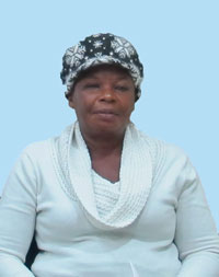
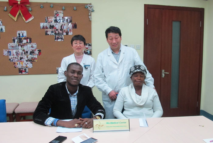
 Name: Taylor Harry Ikoru Ipalibo
Sex: Female
Country: Nigeria
Age: 52 years
Diagnosis: 1. Retinitis Pigmentosa 2. HLP(hyperlipidaemia)
Date: Feb. 2nd, 2015
Days Admitted to Hospital: 21 days
Before treatment:
Four years ago, there was a declined in the patient’s left eye vision with no reason. This was a progressive growth. Several months later, her right eye vision declined. She diagnosed as Retinitis Pigmentosa by Haywards Heath hospital and she was untreated. As it stands now, her left eye can only focus the outline of big objects. Close distinguish fingers: right eye was unable to distinguish fingers at close distance. She wants to have a better treatment, so she came to our hospital and she was diagnosed with retinitis pigmentosa.
She was in good spirit. Her sleep, eating, urination and excrement were good.
Admission PE:
Bp: 140/89mmHg; Hr: 64/min. Br: 19/min. Her nutrition status is normal. Her skin and mucosa color is normal, no yellow stains, petechial or bruise. Pharyngeal hyperemia was obvious with no swollen in bilateral tonsil. The respiratory sounds in both lungs were clear, with no obvious rales. The rhythm of her heartbeat was normal, with no obvious murmur. Her abdomen was soft, with no pressing pain or rebound tenderness. The liver and spleen were normal. Blood cholesterol and triglyceride were high.
Nervous System Examination:
Patient was alert. Her mental status was good, the memory ability and calculation ability are normal. Both pupils were equal in size, the diameter was 3mms, react well to light stimulus. Patient had tunnel vision in left side. The right side is normal by gross check. Left visual acuity: 4.9/100, right visual acuity: 6/100. She had color vision deficit. Ophthalmoscopy check: right retina color is light yellow, there are black osteoid sediment diffused, the border of optic nerve head and macula is not clear. The A/V: 1:3; left retina color is light yellow; there are big amount of black osteoid sediment diffused, more than right side. The optic nerve is clear, without edema. The macula is clear, the A/V: 1:3. Eyeballs can move freely, no nystagmus. The forehead wrinkle pattern was symmetrical. The bilateral nasolabial sulcus was equal in depth. The tongue was at the middle position. Patient teeth are normal. Her neck could move freely. The muscle tone of 4 limbs are normal, muscle power of 4 limbs is at 5 deg. Abdomen reflex is normal, tendon reflex is normal. Sucking reflex and palm-jaw reflex are negative. Bilateral Hoffmann sign are negative. Rossolimo sign of both sides are negative; the Babinski sign of both sides are negative. The deep and superficial sensations are normal. Patient could finish coordinate movement normally. Meningeal irritation sign is negative.
Treatment:
After her admission, patient had complete detail body examinations. Doctors confirmed the diagnosis as: 1. Retinitis Pigmentosa 2. HLP (hyperlipidaemia). We gave patient treatment to nourish the neurons, improve the blood circulation, etc.
Post-treatment:
After the treatment,the vision of left eye is 5.5/100 in three meters standard visual chart while that of right eye is 10/100. The color discrimination is better. The bone spicule-shaped pigment deposits on the bilateral eye fundus have been reduced. The blood circulation is better, the A/V: 2:3.

